BMRB Entry 15139
Click here to enlarge.
PDB ID:
Entry in NMR Restraints Grid
Validation report in NRG-CING
Chem Shift validation: AVS_full, LACS, SPARTA
BMRB Entry DOI: doi:10.13018/BMR15139
MolProbity Validation Chart
NMR-STAR file interactive viewer.
NMR-STAR v3 text file.
XML gzip file.
RDF gzip file.
All files associated with the entry
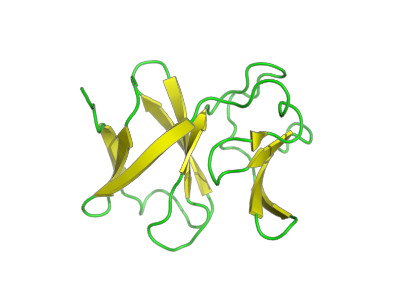
Citation: Ramelot, Theresa; Cort, John; Yee, Adelinda; Arrowsmith, Cheryl; Kennedy, Michael. "NMR structure of E.coli NirD." .
Assembly members:
NirD, polymer, 113 residues, 12750 Da.
Natural source: Common Name: E. coli Taxonomy ID: 562 Superkingdom: Eubacteria Kingdom: not available Genus/species: Escherichia coli
Experimental source: Production method: recombinant technology Host organism: Escherichia coli Vector: pET
Entity Sequences (FASTA):
NirD: QGHMSQWKDICKIDDILPET
GVCALLGDEQVAIFRPYHSD
QVFAISNIDPFFESSVLSRG
LIAEHQGELWVASPLKKQRF
RLSDGLCMEDEQFSVKHYEA
RVKDGVVQLRGGS
- assigned_chemical_shifts
- spectral_peak_list
| Data type | Count |
| 13C chemical shifts | 491 |
| 15N chemical shifts | 119 |
| 1H chemical shifts | 785 |
Additional metadata:
Assembly:
| Entity Assembly ID | Entity Name | Entity ID |
|---|---|---|
| 1 | NirD | 1 |
Entities:
Entity 1, NirD 113 residues - 12750 Da.
Three non-native residues at the N-terminus (QGH), were not included in the calculation. Two non-native residues at the C-terminus (GS), were included in the calculation.
| 1 | GLN | GLY | HIS | MET | SER | GLN | TRP | LYS | ASP | ILE | ||||
| 2 | CYS | LYS | ILE | ASP | ASP | ILE | LEU | PRO | GLU | THR | ||||
| 3 | GLY | VAL | CYS | ALA | LEU | LEU | GLY | ASP | GLU | GLN | ||||
| 4 | VAL | ALA | ILE | PHE | ARG | PRO | TYR | HIS | SER | ASP | ||||
| 5 | GLN | VAL | PHE | ALA | ILE | SER | ASN | ILE | ASP | PRO | ||||
| 6 | PHE | PHE | GLU | SER | SER | VAL | LEU | SER | ARG | GLY | ||||
| 7 | LEU | ILE | ALA | GLU | HIS | GLN | GLY | GLU | LEU | TRP | ||||
| 8 | VAL | ALA | SER | PRO | LEU | LYS | LYS | GLN | ARG | PHE | ||||
| 9 | ARG | LEU | SER | ASP | GLY | LEU | CYS | MET | GLU | ASP | ||||
| 10 | GLU | GLN | PHE | SER | VAL | LYS | HIS | TYR | GLU | ALA | ||||
| 11 | ARG | VAL | LYS | ASP | GLY | VAL | VAL | GLN | LEU | ARG | ||||
| 12 | GLY | GLY | SER |
Samples:
sample_1: protein, [U-100% 13C], 1 ± .2 mM; TRIS 10 ± 1 mM; sodium chloride 300 ± 5 mM; ZINC ION 10 ± 1 uM; DTT 10 ± 1 mM; sodium azide 0.01 ± 0.001 %; H2O 95%; D2O 5%
sample_2: protein, [U-100% 13C], 1 ± .2 mM; TRIS 10 ± 1 mM; sodium chloride 300 ± 5 mM; ZINC ION 10 ± 1 uM; DTT 10 ± 1 mM; sodium azide 0.01 ± 0.001 %; D2O 100%
sample_conditions_1: ionic strength: 300 mM; pH: 7.3; pressure: 1 atm; temperature: 293 K
Experiments:
| Name | Sample | Sample state | Sample conditions |
|---|---|---|---|
| 3D 1H-15N NOESY | sample_1 | isotropic | sample_conditions_1 |
| 2D 1H-15N HSQC (NH2 only) | sample_1 | isotropic | sample_conditions_1 |
| 2D 1H-13C HSQC (aliph) | sample_1 | isotropic | sample_conditions_1 |
| 3D HNCACB | sample_1 | isotropic | sample_conditions_1 |
| 3D 1H-13C NOESY (aliph) | sample_1 | isotropic | sample_conditions_1 |
| 4D CC NOESY | sample_2 | isotropic | sample_conditions_1 |
| 3D 1H-13C NOESY (arom) | sample_1 | isotropic | sample_conditions_1 |
| 2D 1H-15N HSQC (open) | sample_1 | isotropic | sample_conditions_1 |
| 2D 1H-15N HSQC (optimized) | sample_1 | isotropic | sample_conditions_1 |
| 2D 1H-13C HSQC (arom ct) | sample_1 | isotropic | sample_conditions_1 |
Software:
AutoStruct v2.1.1, Huang, Swapana, Rajan, Ke, Xia, Shukla, Inouye and Montelione - data analysis
X-PLOR NIH v2.15.0, Schwieters, Kuszewski, Tjandra and Clore - structure solution
CNS v1.1, Brunger, Adams, Clore, Gros, Nilges and Read - refinement
SPARKY v3.1, Goddard - peak picking
NMRPipe vlinux9, Delaglio, Grzesiek, Vuister, Zhu, Pfeifer and Bax - processing
NMR spectrometers:
- Varian INOVA 600 MHz
- Varian INOVA 750 MHz
Download HSQC peak lists in one of the following formats:
CSV: Backbone
or all simulated peaks
SPARKY: Backbone
or all simulated peaks